Über 7 millionen englischsprachige bücher. The knee joint is a synovial joint which connects the femur. Mri knee anatomy | knee sagittal anatomy | free cross sectional anatomy. Stanford bone tumor ddx | iss/ssr msk lectures | search ocad cases | stanford virtual readouts stanford msk mri atlas has served over 1,000,000 pages to users in over 100 countries. On axial mr images, the semimembranosus muscle is located between the medial head of the gastrocnemius muscle and the gracilis muscle (fig.
Atlas Of Knee Mri Anatomy W Radiology from w-radiology.com Injuries such as anterior cruciate ligament, meniscus and rotator cuff tears are all easily diagnosed when there is a firm understanding and knowledge of human anatomy. Their origins and insertions are difficult to remember, and they are best considered as parts of general functional groups. Anatomy basic knee mri checklist. Knee muscle anatomy mri / jaypeedigital ebook reader. Usually, the images are taken in three planes; T2w axial fat sat 1. Knee mri anatomy of the knee anterior cruciate ligament pet ct journal prompts biceps study health fitness. Anatomical structures of the lower limb (hip, thigh, knee, leg, ankle and foot) and specific regions (compartment of the lower.
Anatomy basic knee mri checklist.
Anatomy arthrogram anatomy basic shoulder mri. An mri of the knee of a healthy subject was performed in the 3 planes of space (coronal, axial, sagittal) commonly used in osteoarticular imaging, with two weightings most commonly used to explore the musculoskeletal pathology of the knee: Please email baodo at stanford.edu Mri knee joint anatomy the knee is placed in 10 to 15 of external rotation esp for sagittal image slicethickness 3 4 mm sections are used for axial coronal and sagittal images of the knee. The knee joint is a synovial joint which connects the femur. The knee joint is a complex structure that involves bones, tendons, ligaments, muscles, and other structures for normal function. Über 7 millionen englischsprachige bücher. Mri knee anatomy scroll using the mouse wheel or the arrows. In this presentation mri anatomy biceps femoris muscle. While a detailed explanation of mri protocols and mr physics is beyond the scope of this text, fast spin echo (fse) mri is most commonly utilized for mri of the knee. Use the mouse scroll wheel to move the images up and down alternatively use the tiny arrows (>>) on both side of the image to move the images.>>) on both side of the image to move the images. T2w axial fat sat 1. Das ist das neue ebay.
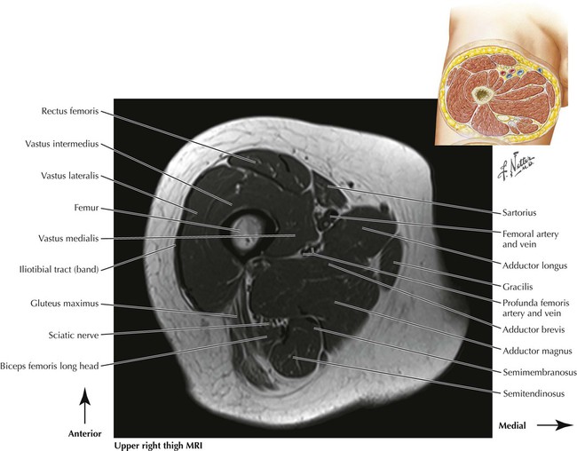
On axial mr images, the semimembranosus muscle is located between the medial head of the gastrocnemius muscle and the gracilis muscle (fig. In this presentation mri anatomy biceps femoris muscle. All mri sections were obtained with a slice thickness of 4 mm, a field of view of 16 cm, and a matrix of 256 in a 1.5. While a detailed explanation of mri protocols and mr physics is beyond the scope of this text, fast spin echo (fse) mri is most commonly utilized for mri of the knee. Anatomical structures of the lower limb (hip, thigh, knee, leg, ankle and foot) and specific regions (compartment of the lower.
X Ray Of The Knee from sinoemedicalassociation.org Anatomical structures of the lower limb (hip, thigh, knee, leg, ankle and foot) and specific regions (compartment of the lower. Ultimately, the image produced by the mri is a thin slice through the knee in one of these three planes. Prescribe sagittal plane off axial images with line parallel to bony glenoid. This mri hip joint axial cross sectional anatomy tool is absolutely free to use. Use the mouse scroll wheel to move the images up and down alternatively use the tiny arrows (>>) on both side of the image to move the images. Iliopsoas psoas major psoas minor iliacus buttocks gluteal r. Anatomy arthrogram anatomy basic shoulder mri. In this presentation mri anatomy biceps femoris muscle.
The muscles of the lower limb are numerous and complex.
This mri hip joint axial cross sectional anatomy tool is absolutely free to use. Mri knee joint anatomy the knee is placed in 10 to 15 of external rotation esp for sagittal image slicethickness 3 4 mm sections are used for axial coronal and sagittal images of the knee. Their origins and insertions are difficult to remember, and they are best considered as parts of general functional groups. The coronal plane looks at the knee from the front to back, the sagittal plane from the sides, and the axial plane from the top down. Use the mouse scroll wheel to move the images up and down alternatively use the tiny arrows (>>) on both side of the image to move the images. Usually, the images are taken in three planes; All mri sections were obtained with a slice thickness of 4 mm, a field of view of 16 cm, and a matrix of 256 in a 1.5. The muscles of the lower limb are numerous and complex. Knee muscle anatomy mri / jaypeedigital ebook reader. Through some of our legacy series, case review, professional and advanced orthopaedic and joint, we round out our discussion of the pathology that occurs in the knee. Knee muscle anatomy axial mri : In this presentation mri anatomy biceps femoris muscle. Three conventional mri planes that are utilized to evaluate the knee include sagittal (oblique), coronal, and transaxial planes.
Three conventional mri planes that are utilized to evaluate the knee include sagittal (oblique), coronal, and transaxial planes. T2w axial fat sat 1. An mri of the knee of a healthy subject was performed in the 3 planes of space (coronal, axial, sagittal) commonly used in osteoarticular imaging, with two weightings most commonly used to explore the musculoskeletal pathology of the knee: The muscles of the lower limb are numerous and complex. T2w axial fat sat 1.
Lower Limbs Radiology Key from radiologykey.com The coronal plane looks at the knee from the front to back, the sagittal plane from the sides, and the axial plane from the top down. This mri knee sagittal cross sectional anatomy tool is absolutely free to use. Mri of the knee is often performed for presumed musculoskeletal conditions. All mri sections were obtained with a slice thickness of 4 mm, a field of view of 16 cm, and a matrix of 256 in a 1.5. Anatomy basic knee mri checklist. While a detailed explanation of mri protocols and mr physics is beyond the scope of this text, fast spin echo (fse) mri is most commonly utilized for mri of the knee. Three conventional mri planes that are utilized to evaluate the knee include sagittal (oblique), coronal, and transaxial planes. Coronal, sagittal and axial plane.
Mri wrist anatomy scroll using the mouse wheel or the arrows.
C m c j o i n t m c p j o i n t i p j o i n t m e t a c a r p a l p r o x i m a l p h a l a n x jun 17, 2021 · knee joint (articulatio genu) the knee joint is. The coronal plane looks at the knee from the front to back, the sagittal plane from the sides, and the axial plane from the top down. Knee muscle anatomy mri / jaypeedigital ebook reader. Knee muscle anatomy axial mri : Über 7 millionen englischsprachige bücher. Positioning for mri upper legs position the patient in supine position with feet pointing towards the magnet feet first supine position the patient over the spine coil and place the body coils over the thighs anterior superior iliac spine down to knee joints. This mri knee sagittal cross sectional anatomy tool is absolutely free to use. Injuries such as anterior cruciate ligament, meniscus and rotator cuff tears are all easily diagnosed when there is a firm understanding and knowledge of human anatomy. In the two most recent series, meniscus mri and mri of the supporting structures, we focus on two important areas of assessment. Knee mri anatomy & diganoses covered in this course Use the mouse scroll wheel to move the images up and down alternatively use the tiny arrows (>>) on both side of the image to move the images.>>) on both side of the image to move the images. In this presentation mri anatomy biceps femoris muscle. Coronal, sagittal and axial plane.


0 Komentar